.png)
How Dermal Clinicians Assess Pigmentation: A Clinical Guide


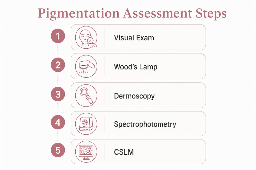
Dermal clinicians assess pigmentation through a layered process that combines visual examination, ultraviolet light analysis, patient history, and instrumental measurement to determine pigment type, depth, and distribution. The goal is never just to name a condition. It is to understand exactly where the pigment sits, what is driving it, and how the skin will respond to treatment. Getting that wrong, even slightly, can mean choosing a laser therapy that worsens vascular redness or applying a peel to dermal pigment that will not budge. Precision here is everything.
The core assessment framework covers:
Visual inspection of pigment hue, border definition, contrast, and distribution pattern
Wood’s lamp examination using UVA light to reveal melanin depth and highlight epidermal versus dermal pigmentation
Patient history review including triggers, duration, medications, and sun exposure history
Dermoscopy for magnified evaluation of pigment morphology and vascularity
Reflectance spectrophotometry using devices like the Konica Minolta CM700D and Delfin SkinColorCatch for quantitative melanin and erythema indices
In vivo confocal scanning laser microscopy for cellular-level, noninvasive subsurface imaging
Standardized visual scales such as the Taylor Hyperpigmentation Scale to reduce subjectivity and track progression across Fitzpatrick skin types I–VI
Each method answers a different clinical question. Used together, they give clinicians the full picture needed to plan treatment with confidence.
How dermal clinicians assess pigmentation with visual and UV examination
Visual assessment is where every pigmentation evaluation begins. A clinician examines the lesion’s color, border sharpness, symmetry, and distribution pattern under good lighting, looking for clues about pigment origin. Brown tones typically suggest epidermal melanin, while gray or blue-brown hues point toward deeper, dermal deposits. The contrast between the lesion and surrounding skin, and whether borders are sharp or diffuse, adds further diagnostic weight.
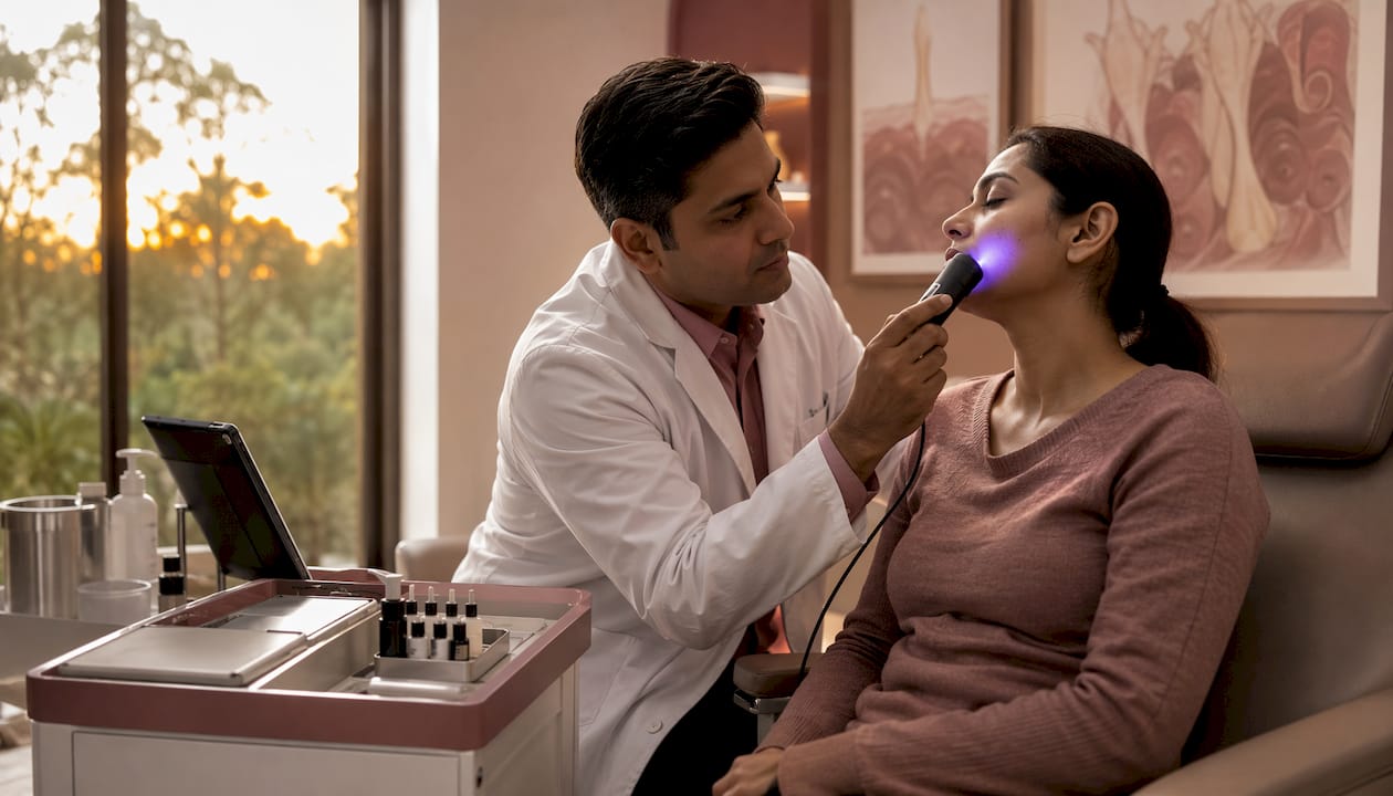
Wood’s lamp examination extends what the naked eye can detect. The lamp emits UVA light to highlight melanin distribution changes, making pigmentary disorders visible that would otherwise blend into the surrounding skin tone. Epidermal pigmentation fluoresces and intensifies under Wood’s lamp, while dermal pigmentation does not enhance, giving clinicians a reliable first-pass indicator of pigment depth. Advanced imaging in pigmentation research confirms that Wood’s lamp combined with multi-mode imaging can reliably distinguish epidermal (brown) from dermal (gray-brown) pigmentation, with direct implications for treatment responsiveness.
Patient history is not a formality. Knowing when a lesion appeared, what triggers it, whether it fluctuates with sun exposure or hormonal changes, and what treatments have been tried before all sharpen the clinical picture considerably. A melasma that worsened during pregnancy tells a different story than one that appeared after a chemical burn, even if the two look similar under standard lighting.
Assessment method | What it reveals | Clinical limitation |
Visual inspection | Pigment hue, border, distribution | Subjective; affected by ambient lighting |
Wood’s lamp | Epidermal vs. dermal depth | Less reliable on Fitzpatrick types V–VI |
Patient history | Triggers, duration, prior treatments | Relies on accurate patient recall |
Combined visual + UV | Depth and pattern confirmation | Requires trained clinical interpretation |
Key clinical signs to evaluate during visual and UV assessment:
Color tone: brown suggests epidermal, gray-brown or blue suggests dermal
Border definition: sharp borders often indicate epidermal origin
Enhancement under Wood’s lamp: intensification confirms epidermal melanin
Distribution pattern: diffuse versus focal, symmetric versus asymmetric
Skin type context: Wood’s lamp is less discriminating on darker Fitzpatrick types
Pro Tip: In patients with Fitzpatrick skin types V or VI, Wood’s lamp results can be ambiguous. Pair UV findings with dermoscopy or confocal microscopy to confirm pigment depth before committing to a treatment plan.
Instrumental tools that give you objective pigmentation data
Visual assessment, even with Wood’s lamp, carries inherent subjectivity. Instrumental techniques address that gap by producing quantifiable, reproducible data that supports clinical decisions and tracks treatment outcomes over time.
Dermoscopy
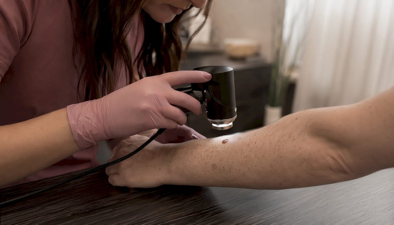
Dermoscopy uses polarized or non-polarized light with magnification to reveal pigment structures invisible to the naked eye. In pigmentation assessment, it allows clinicians to evaluate the morphology of melanin deposits, including their pattern, depth cues, and relationship to vascular structures. Reticular, globular, and homogeneous patterns each carry different diagnostic implications, and dermoscopy is particularly useful in differentiating benign hyperpigmentation from early melanocytic lesions that warrant further investigation. It is widely available, non-invasive, and adds meaningful diagnostic resolution without significant cost.
Reflectance spectrophotometry
Devices like the Konica Minolta CM700D and Delfin SkinColorCatch measure skin color objectively using the CIELAB Lab* color system, which quantifies lightness, redness, and yellowness independently. The L* value tracks overall skin brightness, while the a* value captures erythema (redness from hemoglobin), and b* reflects yellow-brown tones associated with melanin. Spectrophotometry and colorimetry provide objective skin tone measurement using this CIELAB system, quantifying both pigmentation and erythema levels with precision that visual grading cannot match. The practical limitation is cost. These devices are accurate and reproducible, but their price point keeps them largely in research and specialist clinic settings rather than everyday practice.
In vivo confocal scanning laser microscopy
Confocal scanning laser microscopy (CSLM) provides cellular-level imaging to differentiate between pigment depths and types noninvasively, making it particularly valuable in complex diagnostic cases where Wood’s lamp and dermoscopy leave ambiguity. The technology uses near-infrared laser light to produce real-time, high-resolution cross-sectional images of the skin at depths reaching the papillary dermis, without requiring a biopsy. Clinicians can visualize melanocyte distribution, dendritic cell patterns, and the presence of melanin at specific skin layers, all of which inform both diagnosis and prognosis.
Instrument | Primary output | Best clinical use | Key limitation |
Dermoscopy | Pigment morphology and pattern | Differentiating pigment types and vascular lesions | Operator-dependent interpretation |
Konica Minolta CM700D | CIELAB Lab* color values | Quantifying melanin and erythema objectively | High cost; specialist settings |
Delfin SkinColorCatch | Melanin and erythema indices | Monitoring treatment response over time | Less accessible in routine clinics |
Confocal laser microscopy | Subsurface cellular imaging | Complex cases; noninvasive depth confirmation | Expensive; limited clinical availability |
Practical considerations for instrumental assessment:
Dermoscopy is the most accessible and widely adopted instrumental tool in clinical practice
Spectrophotometry excels at tracking subtle color changes across treatment cycles
Confocal microscopy is best reserved for diagnostically ambiguous cases or research contexts
Skincare technology advances continue to bring some of these capabilities into more accessible clinical formats
One point that clinicians must keep front of mind: differentiating melanin from hemoglobin is clinically critical, because treatments targeting melanin can worsen vascular-affiliated pigmentation if the chromophore is misidentified. Spectrophotometry’s ability to separate the a* (erythema) and b* (melanin) channels makes it particularly useful when the clinical picture is ambiguous.
While objective instruments offer high precision, day-to-day pigmentation diagnosis still relies on clinician expertise and patient history as the foundation. Instruments support and confirm; they do not replace clinical judgment.
Why pigment depth diagnosis shapes every treatment decision
Knowing that a patient has hyperpigmentation is only the starting point. Knowing where that pigment sits in the skin determines whether treatment will work, how long it will take, and what risks are involved.
Epidermal pigmentation sits in the upper layers of the skin and typically presents as well-defined, brown lesions. It responds well to chemical peels and light-based therapies, including superficial peels with alpha hydroxy acids and targeted laser treatments. The melanin is accessible, and with the right protocol, results are often visible within a few treatment cycles. Dermal pigmentation is a different challenge. Sitting deeper in the dermis, it tends to appear gray-brown or blue-gray and responds far more slowly and inconsistently to treatment. Aggressive approaches risk post-inflammatory hyperpigmentation, particularly in darker skin types, making cautious, multi-modal protocols the standard of care.
Mixed-type pigmentation, where melanin deposits exist at both epidermal and dermal levels simultaneously, is the most clinically demanding presentation. Treatment must address both layers without over-treating either, which requires precise depth diagnosis before any intervention begins. Skipping that step and defaulting to a single-modality approach often produces partial results or adverse effects.
When to consider biopsy and histopathological confirmation:
Lesions with atypical features under dermoscopy that raise concern for melanocytic pathology
Pigmentation that fails to respond to well-targeted treatment over an appropriate timeframe
Ambiguous Wood’s lamp or confocal findings in a complex clinical presentation
Any lesion where the differential diagnosis includes a condition requiring histological confirmation
Clinical practice guidelines consistently emphasize depth diagnosis as the critical step before selecting any treatment modality. A superficial pigmentation treatment applied to dermal pigment will not penetrate far enough to be effective, while an aggressive laser protocol aimed at epidermal pigment in a patient with mixed-type involvement can trigger a rebound response. The depth diagnosis is not a preliminary step. It is the treatment decision.
Pro Tip: When assessing mixed-type pigmentation, use Wood’s lamp to establish a baseline depth indicator, then confirm with dermoscopy or confocal imaging before selecting your treatment protocol. Document findings at each stage to track how the dermal component responds over time.
How standardized scales and clinical guidelines improve diagnostic consistency
Subjective visual assessment, even by experienced clinicians, varies between practitioners and across clinical settings. Standardized tools exist precisely to reduce that variability, and the evidence for their value in routine practice is strong.
The Taylor Hyperpigmentation Scale uses 15 colored cards to standardize the assessment of hyperpigmentation progression across Fitzpatrick skin types I through VI. Each card represents a calibrated shade, allowing clinicians to match and grade lesion color against a validated reference rather than relying on memory or subjective description. The scale is validated, inexpensive, and practical in both clinical and research settings, making it one of the most accessible standardized tools available for monitoring pigmentation changes reliably over time.
The Fitzpatrick Skin Phototype Scale, while foundational, has well-documented limitations in diverse populations. It was originally designed to predict UV sensitivity rather than to characterize skin color comprehensively, and it tends to compress the range of darker skin tones into fewer categories than the clinical reality warrants. Clinicians working with patients across a broad spectrum of skin tones increasingly supplement Fitzpatrick typing with tools like the Taylor scale or the Individual Typology Angle to capture pigmentation characteristics more accurately.
Standardized visual scales help reduce racial and ethnic diagnostic disparities and improve reliability in pigmentation assessment among diverse skin types. The Skin of Color Society’s clinical guidelines note that assessment must account for genetics, social factors, and cutaneous characteristics, and that combining visual scales with clinical expertise minimizes diagnosis disparities, particularly for patients with skin of color. This matters because underdiagnosis and misdiagnosis of pigmentation disorders in darker skin types remain a documented problem in dermatology.
How to integrate standardized scales into clinical practice:
Use the Taylor Hyperpigmentation Scale at baseline and at each follow-up to track progression objectively
Document Fitzpatrick skin type at intake, and note its limitations when working with types IV–VI
Supplement phototype classification with colorimetric data when spectrophotometry is available
Incorporate patient history alongside scale findings to contextualize triggers and treatment response
Apply skin pigmentation best practices from current clinical guidelines to reduce inter-clinician variability
The Taylor Hyperpigmentation Scale bridges the gap between subjective visual assessment and quantifiable results, proving its value in both clinical trials and routine monitoring. When paired with a thorough patient history and the right instrumental tools, standardized scales give clinicians a repeatable, defensible framework for tracking pigmentation from first assessment through to treatment completion. That consistency is what separates a well-documented clinical outcome from an anecdotal one.
Putting it all together: assessment that leads to confident treatment
Fundamentalskin’s approach to pigmentation reflects exactly this clinical framework. Led by Chevonne, a Dermal Clinician with 15 years of experience, the practice integrates thorough assessment, including skin type evaluation, pigment depth analysis, and patient history, before recommending any treatment. That foundation is what makes targeted treatments like the Larimedical Biomimetic Peel effective: the protocol is chosen because the assessment confirmed the pigment is accessible and the skin is ready.

If you are ready to address pigmentation with a treatment plan built on accurate clinical assessment, the Biomimetic Peel + LED Therapy at Fundamentalskin offers a non-invasive, results-driven option designed for real skin, real concerns, and real outcomes.
Key Takeaways
Accurate pigmentation assessment requires combining visual examination, Wood’s lamp analysis, instrumental measurement, and standardized scales to determine pigment type, depth, and distribution before any treatment is selected.
Point | Details |
Depth diagnosis drives treatment | Epidermal pigment responds to peels and light therapy; dermal pigment requires cautious, multi-modal protocols. |
Wood’s lamp reveals pigment depth | UVA illumination distinguishes epidermal from dermal pigmentation, guiding initial treatment direction. |
Taylor scale reduces subjectivity | The 15-card Taylor Hyperpigmentation Scale standardizes grading across Fitzpatrick types I–VI for consistent monitoring. |
Chromophore differentiation prevents errors | Separating melanin from hemoglobin is critical; misidentification can cause laser treatments to worsen vascular redness. |
Instruments support, not replace, clinical judgment | Spectrophotometry and confocal microscopy add precision, but clinician expertise and patient history remain the diagnostic foundation. |
Recommended
Comments