.png)
How to Identify Pigmentation Types on Skin
- chevonne stewart
- 1 day ago
- 8 min read


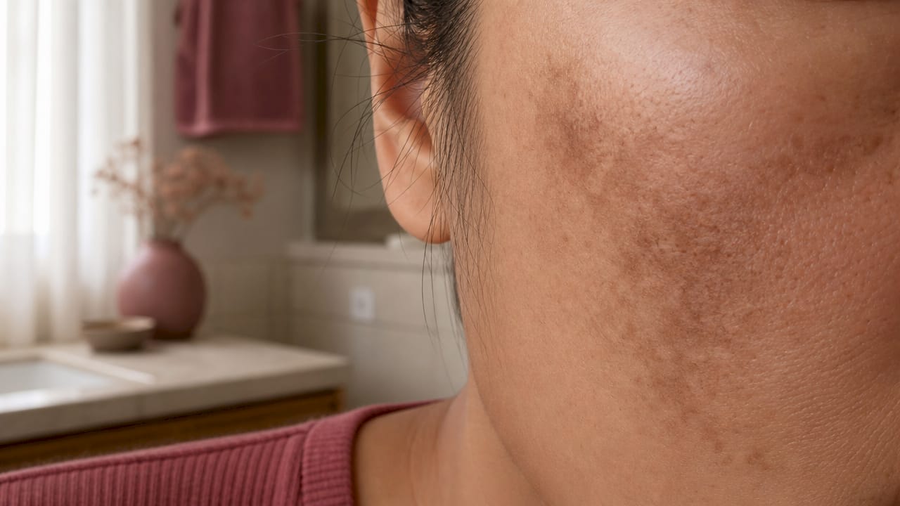
Skin pigmentation is defined as a change in skin color caused by variation in melanin production, the pigment made by cells called melanocytes. To identify pigmentation types on skin accurately, you need to understand three core categories: hyperpigmentation (too much melanin), hypopigmentation (too little melanin), and depigmentation (complete melanin loss, as seen in vitiligo). Skin color changes follow this melanin framework regardless of skin tone. Getting this classification right matters because each type responds to completely different treatments. Treating hyperpigmentation the same way you treat vitiligo, for example, can make both conditions worse.
What are the common types of skin pigmentation?
The three main categories of skin pigmentation each have distinct visual features. Learning those features is the fastest way to narrow down what you are looking at on your own skin.
Hyperpigmentation: darker patches
Hyperpigmentation is the most common category. It appears as patches or spots darker than your natural skin tone. The four most frequently seen types are:
Melasma: Brown or gray-brown patches that appear symmetrically, most often on the cheeks, forehead, upper lip, and chin. Hormones and sun exposure are the primary triggers. Melasma is especially common during pregnancy and while taking oral contraceptives.
Post-inflammatory hyperpigmentation (PIH): Dark marks left behind after acne, eczema, a cut, or any skin injury. PIH follows the shape of the original wound or breakout and appears where the inflammation occurred. Epidermal PIH often resolves in 6–12 months, but deeper pigmentation can persist for years.
Sunspots (solar lentigines): Flat, well-defined brown spots caused by cumulative UV exposure. They appear most often on the face, hands, and shoulders. Unlike freckles, sunspots do not fade in winter.
Freckles (ephelides): Small, flat, light brown spots that are genetically determined and become more visible with sun exposure. They fade significantly in winter and are more common in fair-skinned individuals.
Pro Tip: Look for symmetry. Melasma almost always appears on both sides of the face in a mirrored pattern. PIH is asymmetrical and follows the exact location of a past skin injury.
Hypopigmentation and depigmentation: lighter patches
Hypopigmentation produces patches lighter than your surrounding skin. Depigmentation means the skin has lost all color in that area. Vitiligo is the clearest example of depigmentation. It creates bright white patches with sharp, well-defined borders. Pityriasis alba, by contrast, produces faint, slightly scaly light patches, most often on the face and arms of children and young adults. The borders of pityriasis alba are blurry, not sharp. That border distinction alone can help you tell the two apart before seeing a clinician.
How do professionals diagnose and classify pigmentation types?
Clinical evaluation is the primary method for diagnosing pigmentation conditions like melasma, vitiligo, and PIH. A clinician examines the color, shape, distribution, and border of each patch, then asks about onset, triggers, medications, and family history.
Beyond the visual exam, professionals use several tools:
Wood’s lamp: A handheld UV light that makes pigment differences more visible. It helps differentiate hypopigmented from depigmented skin in vitiligo assessment. However, Wood’s lamp findings are variable by skin phototype and should not be the sole basis for a melasma diagnosis, especially in darker skin tones.
Dermoscopy: A magnifying device that reveals pigment network patterns and vascular features invisible to the naked eye. Dermoscopic features like a brown reticular pigment network and telangiectasias help classify melasma more accurately than Wood’s lamp alone.
Biopsy and mycologic tests: Used when a diagnosis is unclear. Depigmented patches that resemble vitiligo can be caused by fungal infections or inflammatory conditions. Biopsy or mycology tests confirm or rule out these look-alikes.
Blood tests: Ordered when systemic causes are suspected, such as autoimmune conditions associated with vitiligo.
Pro Tip: Do not rely on a UV flashlight at home to confirm your pigmentation type. Wood’s lamp results vary significantly based on skin tone, and a negative result does not rule out a condition.
Step-by-step guide to self-identify your pigmentation type
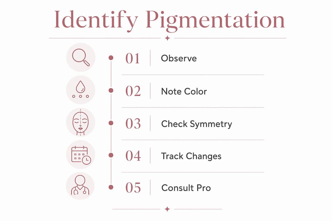
Self-identification is not a replacement for clinical diagnosis. It is a starting point that helps you describe your skin accurately and make better decisions about seeking care. Follow these five steps.
Observe color, shape, and distribution. Stand in natural daylight and look at the affected area. Is the patch darker or lighter than your surrounding skin? Is it flat or raised? Does it appear on one side of your face or both? Symmetrical distribution points toward melasma. A single irregular patch points toward PIH or a sunspot.
Note recent triggers. Think back over the past 3–6 months. Did you have a breakout, a rash, or a skin injury in that exact location? That history strongly suggests PIH. Did the patch appear or worsen after sun exposure? That points toward sunspots or melasma. Hormonal changes, new medications, and pregnancy are also key triggers to note.
Check the borders. Sharp, clearly defined borders are typical of vitiligo and sunspots. Blurry or gradual borders are more common in melasma, PIH, and pityriasis alba. Border clarity is one of the most reliable visual clues available without clinical tools.
Compare to the characteristics table below.
Pigmentation type | Color | Border | Common location | Key trigger |
Melasma | Brown or gray-brown | Irregular but symmetrical | Cheeks, forehead, upper lip | Hormones, UV exposure |
PIH | Brown, red, or purple | Follows injury shape | Anywhere skin was inflamed | Acne, eczema, trauma |
Sunspots | Light to dark brown | Well-defined | Face, hands, shoulders | Cumulative UV exposure |
Freckles | Light brown | Soft | Nose, cheeks | Genetics, UV exposure |
Vitiligo | White | Sharp, well-defined | Face, hands, genitals | Autoimmune |
Pityriasis alba | Pale, slightly pink | Blurry | Face, arms | Eczema, dry skin |
Seek professional evaluation when needed. If a patch is new, spreading quickly, changing shape, or you are unsure after comparing your findings, book an appointment with a dermal clinician. Unexplained pigmentation changes warrant clinical tests including Wood’s lamp, dermoscopy, or blood work depending on the suspected cause.
Pro Tip: Photograph the area in consistent lighting every two weeks. Progress photos help a clinician assess whether the patch is stable, fading, or spreading, which is information that memory alone cannot provide.
Common mistakes when identifying skin pigmentation types
Misidentification is common, and it leads to treatments that either do nothing or actively worsen the condition. These are the most frequent errors.
Confusing vitiligo with pityriasis alba. Both produce lighter patches on the face. Vitiligo patches are bright white with sharp borders. Pityriasis alba patches are pale and slightly scaly with soft edges. Ruling out similar hypopigmented disorders matters because their treatments and prognoses differ significantly.
Mistaking melasma for PIH. Both appear as brown patches on the face. The difference is symmetry and trigger. Melasma is bilateral and hormone-driven. PIH is unilateral and follows a specific injury or breakout.
Treating PIH while inflammation is still active. Treating PIH without addressing the underlying inflammation first can worsen pigmentation. The inflammation must be resolved before pigment-targeting treatments can work.
Assuming all white patches are vitiligo. Fungal infections like tinea versicolor and inflammatory conditions can produce depigmented-looking patches. These require antifungal treatment, not vitiligo protocols.
Skipping sun protection during self-assessment. UV exposure worsens nearly every pigmentation condition. Wearing SPF 30 or higher daily is mandatory while you are observing and assessing your skin.
Skin tone affects how pigmentation appears visually. Darker skin tones may show PIH as deep brown or purple patches rather than the lighter brown seen in fair skin. This variation makes self-identification harder and professional evaluation more valuable.
Rapid spread, irregular borders that keep changing, or pigmentation accompanied by itching, bleeding, or crusting all require prompt medical evaluation. These features can indicate conditions beyond cosmetic pigmentation concerns.
Key Takeaways
Accurate skin pigmentation classification depends on observing color, border, distribution, and triggers together, not on any single visual feature alone.
Point | Details |
Three core categories | Hyperpigmentation, hypopigmentation, and depigmentation each have distinct visual features and causes. |
Symmetry is a key clue | Melasma appears symmetrically on both sides of the face; PIH follows the shape of a past injury. |
Wood’s lamp has limits | UV lamp results vary by skin tone and should not be the sole basis for identifying melasma. |
Treat inflammation first | PIH will not resolve if the underlying inflammation is still active; photoprotection is mandatory. |
Professional evaluation matters | New, spreading, or unusual patches require clinical assessment including dermoscopy or biopsy. |
What 15 years in a treatment room taught me about pigmentation
Most people arrive at a skin clinic having already treated themselves for the wrong condition. That is not a criticism. It is genuinely hard to tell melasma from PIH when you are looking at your own face in a bathroom mirror. What I have learned over 15 years working with clients on pigmentation is that the trigger history is almost always more revealing than the patch itself.
When a client tells me a dark patch appeared two months after a breakout in that exact spot, I already have a strong working picture before I even examine the skin. When another tells me her patches appeared during her second pregnancy and sit symmetrically across both cheeks, that is a different conversation entirely. The patch looks similar. The cause and the treatment are completely different.
Self-identification is genuinely useful. It helps you walk into a consultation with context, ask better questions, and understand the reasoning behind a treatment plan. What it cannot do is replace dermoscopy, a trained eye, or a biopsy when one is needed. The goal of learning to read your own skin is not to diagnose yourself. It is to become an informed participant in your own care. That shift alone changes outcomes. Clients who understand their pigmentation type follow photoprotection advice more consistently, and that consistency is what separates a result that lasts from one that fades within months.
Photoprotection is not optional during any stage of pigmentation assessment or treatment. UV exposure worsens every category of pigmentation. Checking your post-treatment skin protection routine is as important as identifying the condition in the first place.
— chevonne
Pigmentation treatments available at Fundamentalskin
Once you have a clearer picture of your pigmentation type, the next step is choosing a treatment matched to that specific condition.

At Fundamentalskin, Chevonne offers a range of targeted, non-invasive treatments designed for women dealing with pigmentation concerns. The Larimedical Biomimetic Peel addresses pigmentation at the cellular level without downtime, making it a strong option for melasma, PIH, and sunspots. The Biomimetic Peel combined with LED therapy supports pigmentation correction while calming inflammation, which is particularly relevant for PIH. The Synergie Peel offers a customized protocol suited to different pigmentation depths and skin tones. Every treatment plan at Fundamentalskin begins with a professional skin assessment so the right approach is matched to your specific pigmentation type.
FAQ
What are the three main types of skin pigmentation?
The three main types are hyperpigmentation (excess melanin causing dark patches), hypopigmentation (reduced melanin causing light patches), and depigmentation (complete melanin loss causing white patches, as in vitiligo).
How do I tell melasma apart from post-inflammatory hyperpigmentation?
Melasma appears symmetrically on both sides of the face and is triggered by hormones or UV exposure. PIH appears where a specific skin injury or breakout occurred and is asymmetrical.
Is Wood’s lamp reliable for identifying pigmentation at home?
Wood’s lamp results vary significantly based on skin tone and should not guide treatment decisions on their own. Clinical evaluation by a trained professional gives a more accurate result.
How long does post-inflammatory hyperpigmentation last?
Epidermal PIH typically fades in 6–12 months with consistent sun protection. Deeper pigmentation can persist for years without targeted treatment.
When should I see a professional about a pigmentation change?
See a clinician if a patch is new, spreading, changing shape, or accompanied by itching, bleeding, or crusting. These features require clinical assessment beyond self-identification.
Recommended
Comments